
Electrotherapy
We pride ourselves on delivering treatment protocols which have clinical research study support. Whilst many Physiotherapy Centre’s use electrotherapy as their main source of treatment delivery, here at BPRC we use electrotherapy treatment parameters sparingly. While some people find electrotherapy helpful, others do not. In addition, medical literature on electrotherapy’s effectiveness has been mixed, and not all electrotherapy treatments are supported by research.
Taking this information into account, we will only use electrotherapy in conjunction with other modalities if there is sufficient clinical evidence to support a treatment protocol of this type.
We have a few Electrotherapy modalities which we administer and even though Ultrasound does not specifically come under this form of treatment, we will list it in this section due to its close links with this type of treatment delivery.
Electrical Muscle Stimulation (EMS)
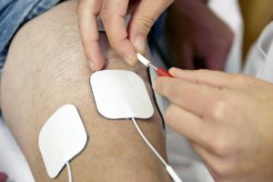 Also known as neuromuscular electrical stimulation (NMES) or electromyostimulation, is the elicitation of muscle contraction using electric impulses. The impulses are generated by a device and delivered through electrodes on the skin in direct proximity to the muscles to be stimulated. The impulses mimic the action potential coming from the central nervous system, causing the muscles to contract. The electrodes are generally pads that adhere to the skin. EMS is both a form of electrotherapy and of muscle training. It is cited by important authors as complementary technique for sport training, and there is published research on the results obtained.
EMS is used for relaxation of muscle spasms, prevention and retardation of disuse atrophy, increase of local blood circulation, muscle rehabilitation and re-education electrical muscle stimulation, maintaining and increasing range of motion, management of chronic and intractable pain, post-traumatic acute pain, post-surgical acute pain, immediate post-surgical stimulation of muscles to prevent venous thrombosis, wound healing and drug delivery.
EMS causes adaptation, i.e. training, of muscle fibres. Because of the characteristics of skeletal muscle fibres, different types of fibres can be activated to differing degrees by different types of EMS, and the modifications induced depend on the pattern of EMS activity. These patterns, referred to as protocols or programs, will cause a different response from contraction of different fibre types. Some programs will improve fatigue resistance, i.e. endurance, others will increase force production. EMS can be used both as a training and a therapeutic tool.
In medicine EMS is used for rehabilitation purposes in Physiotherapy in the prevention of disuse muscle atrophy which can occur for example after musculoskeletal injuries, damage to bones, joints, muscles, ligaments and tendons.
Interferential Therapy
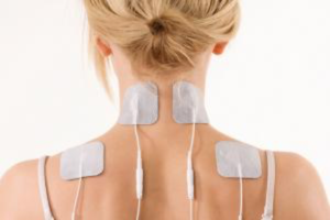 Interferential current (IF) therapy, or interferential stimulation, is a more complex development of TENS, and is a unique way of effectively delivering therapeutic frequencies to tissue.
While conventional TENS devices deliver electrical pulses at extremely low frequency (typically 2-150 Hz), Interferential stimulation uses medium frequencies — above 1000 Hz, and typically around 4000 Hz. Medium frequencies travel in the tissue much more easily, can go further and deeper,and with less discomfort to the patient.
Interferential is so named because “interference” between currents of multiple frequencies is what makes interferential devices effective. The “interference” occurs between the 2 or more currents used. One current has fixed frequency (typically at 4000 Hz) and the other’s frequency varies by up to 400 Hz. At the point of intersection between the electrodes (which can be deeper than in TENS due to the medium current’s ease of penetration) the combined currents produce an “interference frequency” also called a “beat” which is a TENS-like low frequency. For example 100 Hz, and for body tissue it has a similar pain-relieving effect to TENS. As well as greater depth, interferential also allows an increased dosage because of the body tissue’s better tolerance of medium-frequency currents. By using four electrodes Interferential treatment (IF) allows better focus and even deeper tissue stimulation. A more detailed discussion of interferential follows.
Conventional TENS and Neuromuscular stimulators use discrete electrical pulses delivered at low frequencies of 2-160 Hz per second. However, Interferential stimulators use a fixed carrier frequency of 4,000 Hz per second and a second adjustable frequency of 4,001-4,400 Hz per second. When the fixed and adjustable frequencies combine (heterodyne), they produce the desired signal frequency (Interference frequency, or beat). Interferential stimulation is concentrated at the point of intersection between the electrodes. This concentration occurs deep in the tissues as well as at the surface of the skin. Conventional TENS and neuromuscular stimulators deliver most of the stimulation directly under the electrodes. Thus, with interferential stimulators, current perfuses to greater depths and over a larger volume of tissue than most forms of electrotherapy. When current is applied to the skin, capacitive skin resistance decreases as pulse frequency increases.’ For example, at a frequency of 4,000 Hz (interferential unit) capacitive skin resistance is eighty (80) times lower than with a frequency of 50 Hz (in the TENS range). Thus, interferential current crosses the skin with greater ease and with less stimulation of cutaneous nociceptors allowing greater patient comfort during electrical stimulation. In addition, because medium-frequency (interferential) current is tolerated better by the skin, the dosage can be increased, thus improving the ability of the Interferential current to permeate tissues and allowing easier access to deep structures. This explains why Interferential current may be most suitable for treating patients with deep pain, for promoting osteogenesis in delayed and non-union fractures, stimulating deep skeletal muscle to augment the muscle pump mechanism in venous insufficiency and for depressing the activity of certain cervical and lumbosacral sympathetic ganglia in patients with increased arterial constrictor tone.
Ultrasound
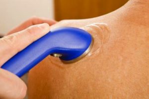 Ultrasonic waves or sound waves of a high frequency that is not audible to the human ear are produced by means of mechanical vibration in the metal treatment head of the ultrasound machine. The treatment head is then moved over the surface of the skin in the region of the injury transmitting the energy into the tissues.
When sound waves meet air it causes a dissipation of the waves, and therefore a special ultrasound gel is placed on the skin to ensure maximal contact between the treatment head and the surface of the skin to provide a medium through with the sound waves can travel. Ultrasound can also be applied under water which is also a medium for ultrasound waves to travel through.
The effects of therapeutic ultrasound are still being disputed. To date, there is still very little evidence to explain how ultrasound causes a therapeutic effect in injured tissue. Nevertheless practitioners world-wide continue to use this treatment modality relying on personal experience rather than scientific evidence. Below are a few theories by which ultrasound is proposed to cause a therapeutic effect.
As the ultrasound waves pass from the treatment head into the skin they cause the vibration of the surrounding tissues, particularly those that contain collagen. This increased vibration leads to the production of heat within the tissue. In most cases this cannot be felt by the patient themselves. This increase in temperature may cause an increase in the extensibility of structures such as ligaments, tendons, scar tissue and fibrous joint capsules. In addition, heating may also help to reduce pain and muscle spasm and promote the healing process.
One of the greatest proposed benefits of ultrasound therapy is that it is thought to reduce the healing time of certain soft tissue injuries.
Ultrasound is thought to accelerate the normal resolution time of the inflammatory process by attracting more mast cells to the site of injury. This may cause an increase in blood flow which can be beneficial in the sub-acute phase of tissue injury. As blood flow may be increased it is not advised to use ultrasound immediately after injury.
Ultrasound may also stimulate the production of more collagen which is the main protein component in soft tissue such as tendons and ligaments. Hence, ultrasound may accelerate the proliferative phase of tissue healing. It is thought to improve the extensibility of mature collagen and so can have a positive effect to on fibrous scar tissue which may form after an injury.
Ultrasound is normally applied by use of a small metal treatment head which emits the ultrasonic beam. This is moved continuously over the skin for approximately 3-5 mins. Treatments may be repeated 1-2 times daily in more acute injuries and less frequently in chronic cases.
Ultrasound dosage can be varied either in intensity or frequency of the ultrasound beam. Simply speaking lower frequency application provides a greater depth of penetration and so is used in cases where the injured tissue is suspected to be deeply situated. Conversely, higher frequency doses are used for structures that are closer to the surface of skin.
For further details please contact our Client Care Team.
Telephone: 00 973 35193767
Email: [email protected]